The knee is the largest and most complex joint in the body, containing numerous parts that have to work in concert for proper joint function. When identifying the source of knee pain or dysfunction, it’s critical to begin with a strong foundational knowledge of knee anatomy and the functions of the joint’s components. We will provide a basic overview of the knee anatomy to help you understand more about your knee and focus your understanding of your own knee problem, to help you discuss your knee situation during your visit with Dr. Buechel.
Bones
 The bones of the knee are the femur, or thigh bone. The tibia, or shin bone. The fibula, the little long bone on the outside of the leg. The patella, or knee cap, that is in the front of the knee and is located inside the tendon of the quadriceps and patella tendon. Bones provide the structural support of the body and where bones connect and motion occurs we call this a joint.
Cartilage
The white, smooth, surface coating on the ends of the bones where two bones meet and move is called cartilage. This cartilage is made up of many cell layers oriented in different directions that provide its different functions. This surface provides painless smooth motion, resistance to shear forces, and shock absorption between the bone ends.
Knee Joint
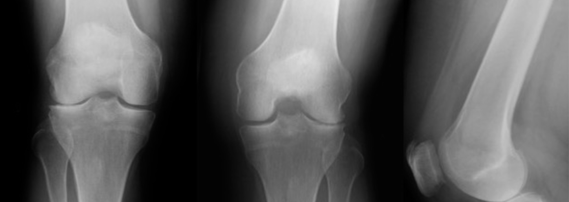
The knee joint is the location where the end of the thigh bone and the top of the shin bone and fibula meet. All of the structures in this location make up the knee joint. The knee joint moves smoothly in healthy knees because of the coating on the end of the bones called cartilage. The Knee joint has three main compartments.
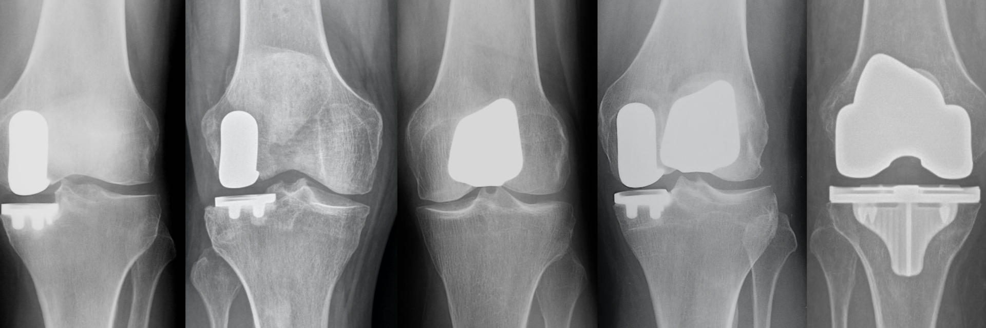
- The Medial Compartment is the inner side of the knee joint between the thigh bone (femur) and shin bone (tibia). Medial compartment is the most common compartment that wears down in the knee and the most common location for partial knee replacement. Partial knee candidates have pain with weight-bearing in just the medial compartment.
- The Lateral Compartment is on the outer side of the knee joint between the thigh bone (femur) and shin bone (tibia). The lateral compartment is the second most common compartment that wears down in the knee. Lateral compartment partial knee candidates have pain with weight-bearing in just the lateral compartment.
- The Patellofemoral Compartment is the third compartment of the knee. This is the compartment in the front of the knee between the kneecap (patella) and the thigh bone (femur). The patella moves down the middle of the two knuckles (condyles) on the end of the femur and increases the efficiency of the quadriceps muscle. Patients with pain in the patellofemoral compartment often describe pain with rising and sitting, stair climbing and knee bending. Patellofemoral partial knee candidates have pain in the anterior compartment under the kneecap (the front of the knee), but no pain in the medial or lateral joint compartments.
Tendons
A tendon is a band of strong whitish connective tissue that connects muscles to bone. These are easy to see on the back of your hand when you move your fingers and the back of your heel where you can see and feel your Achilles’ tendon. Two important tendons located in the front of the knee are the quadriceps tendon and the patellar tendon. The quadriceps tendon connects the patella, or kneecap, to the quadriceps muscle, located on the front of the thigh. The patellar tendon connects the patella to the tibia, or shinbone. Because the patella tendon actually connects two bones, the patellar tendon is technically a ligament. This system on the front of the knee is called the “extensor mechanism” because when you contract your quadriceps muscle it pulls on the quadriceps tendon, patella, and patella tendon which pulls your shin bone up, extending your knee.
Ligaments
Ligaments are the strong, rope-like tissues that connect bones to bones, preventing unnecessary motion and maintaining joint stability. The knee contains four ligaments:
- The ACL (anterior cruciate ligament), is located in the center of the knee. This ligament connects the end of the femur to the top front of the tibia and limits the forward movement of the tibia.
- The PCL (posterior cruciate ligament), is also located in the center of the knee. This ligament connects the end of the femur to the the back center of the tibia and limits the backward movement of the tibia.
- The MCL (medial collateral ligament), on the inner side of the knee is connected to the inner side of the femur and inner side of the tibia and it prevents the leg from moving too far to the outside, stabilizing the inner side of the knee.
- The LCL (lateral collateral ligament), on the outer side of the knee is connected to the outside of the femur and outside of the knee at the fibula and it prevents the leg from moving too far to the inside, stabilizing the outer side of the knee.
Muscles
The hamstrings and quadriceps muscles in the back and front of the thigh and knee joint provide the leg with the ability to flex and extend the knee joint. The quadriceps are four muscles in the front of the thigh that connect to the patella via the quadriceps tendon which assists with extending the knee. The hamstrings, a muscle group in the back of the thigh connect to the back of the tibia at the knee level and serve to flex, or bend the knee joint. The gluteal muscles, located higher up on your backside, serve to stabilize and move the pelvis and upper leg. The calf muscles in the back of the knee (gastroc and soleous muscles) connect the back end of the femur to the heel bone with the Achilles tendon.
Joint Capsule
The knee joint is enclosed by the joint capsule, a strong connective tissue membrane like a ballon around the knee joint that contains the joint fluid (synovial fluid), a liquid found in the body’s mobile joints. This fluid provides lubrication and nutrients to the joint. In a healthy joint, only a few drops are needed to lubricate the healthy cartilage surfaces to maintain smooth motion.
Bursa
Knee bursa are little potential fluid pockets that surround the knee joint and are usually located where tendons connect to the bones to help act as a protective cushion between the bone and tendon insertion. In pathologic situations these bursa can get inflamed and swollen causing pain at their location.
Meniscus
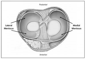
The meniscus is a tough, smooth, rubbery C-shaped piece of cartilage (fibrocartilage) that is wedged shaped in profile, and sits between the cartilage surfaces of the bones. It distributes your body weight more evenly across the knee joint and improves the stability of the joint. Each knee has two menisci, the medial (inside) and the lateral (outside). They are physically attached to the top of the shin bone (tibia) and make contact with the thigh bone (femur). They act as shock absorbers during weight-bearing activities.
- The front ⅓ is called the “anterior horn”.
- The middle ⅓ is called the “body”.
- The back ⅓ is called the “posterior horn”.
The wedged shaped profile helps maintain the stability of the joint by keeping the rounded femur surface from sliding off the flat tibial surface. The meniscus is nourished by small blood vessels coming in from the outer edges (vascular zone, or red zone). The meniscus also has a large area in the inner ⅓ that has no direct blood supply (avascular zone, or white zone). Between these zones is an area where the blood supply is minimal, called the red/white zone.
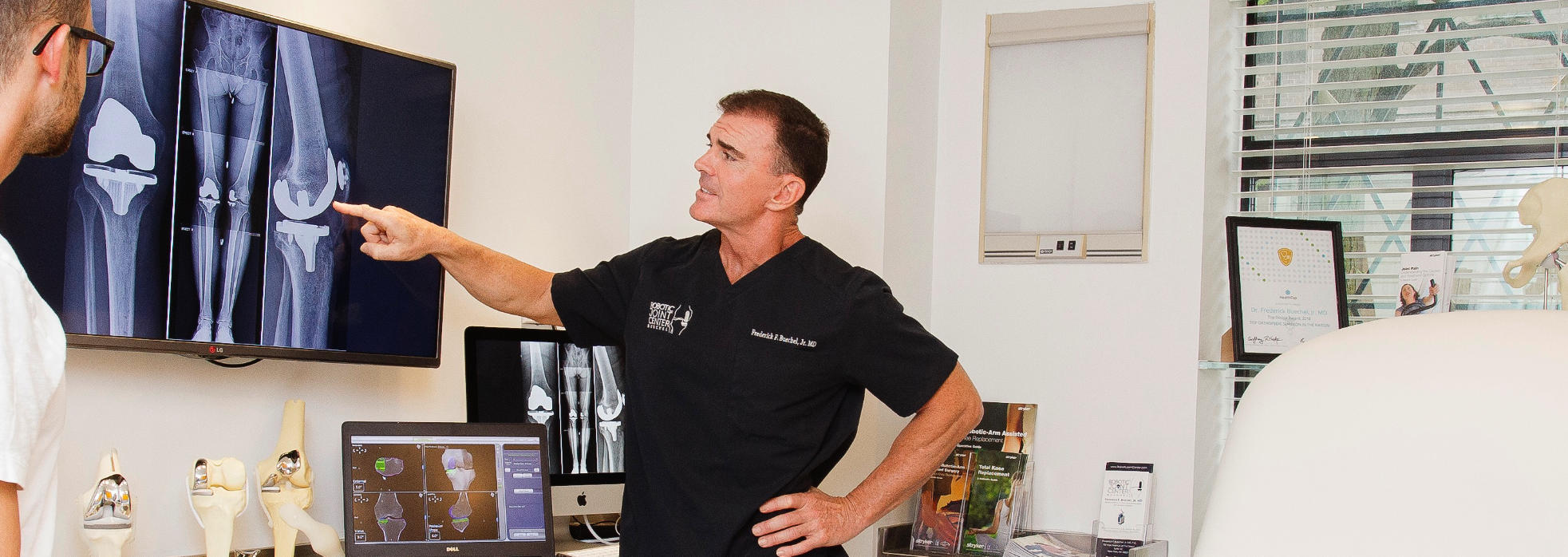
Written by: Dr. Frederick Buechel, Jr.
Dr. Buechel is a Stryker certified international instructor and an expert in the field of Robotic-Arm Assisted Knee & Hip Replacement.
Dr. Buechel performs Outpatient Robotic Partial Knee replacement surgery, and Outpatient or short stay Robotic Total Knee replacement in New York City at Lenox Hill Hospital, Midtown Surgery Center and New York Center for Ambulatory Surgery in Manhattan. Dr. Buechel operates at St. Barnabas Medical Center Hospital in Livingston, New Jersey. Internationally Dr. Buechel performs Robotic Knee and Hip replacement in Taipei, Taiwan at Taipei Postal Hospital.
Meet Dr. Buechel