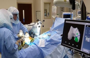
Dr. Buechel’s Mako™ Robotic Partial Knee Replacement Operating Room Setup
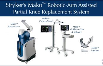
Mako™ Robotic Partial Knee Replacement System used by Dr. Buechel
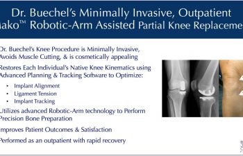
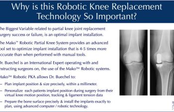
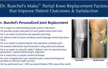
Mako™ Robotic-Arm Assisted Partial Knee Surgery is the most consistently, reproducible, precision joint replacement installation system available in the world today. Dr. Buechel is an expert user of this Mako™ system. He understands which patients are best suited for this procedure and he has an international surgical experience of over 1,000 robotic procedures, successfully operating on people from all over the world. This vast experience allows him to optimize each person’s knee implant installation, which improves the implants function & longevity, while creating the most “natural feel” for the patient.
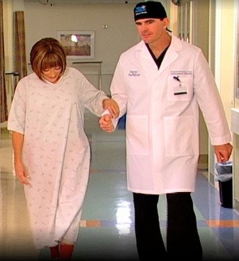
After their procedures, Dr. Buechel’s patients are walking the day of surgery. Most patients are off prescription pain medications within days, and they feel better than before surgery often in just 2-4 weeks. This rapid return to function is the result of Dr. Buechel’s attention to every detail, and meticulous management of the knee and surrounding tissues during the procedures.
Patients will often start their process with a complimentary online consultation prior to proceeding with their office visit. This process is ideal for those out of the area that will eventually be traveling to see Dr. Buechel for their office visit and scheduled procedure. Prior to the procedure, patients are seen for their in person, comprehensive office visit. Once confirmed in the office that a patient is a good candidate & properly indicated for the procedure, their preoperative planning CT scan is obtained and the procedure is confirmed as scheduled.
Video of Robotic Partial Knee Replacement Surgery Process and Procedure
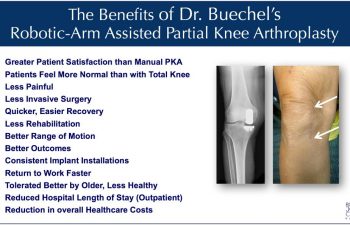
The Benefits of Dr. Buechel’s Mako™ Robotic Partial Knee Replacement
The benefits of using the Mako™ Robotic-Arm System when performing partial knee replacement over conventional manual tool systems has now been well documented in the peer review literature, of which Dr. Buechel has contributed greatly from his clinical experiences. As a world leading surgeon expert and an educator of surgeons with this technology, Dr. Buechel has seen first-hand, all of the benefits patients have enjoyed over a decade of involvement with Mako™ Robotics. If your knee is painful, and you are considering joint replacement surgery, you should contact Dr. Buechel through the office, or our initial Complimentary Online Consultation, to see if you can benefit, also.
The Process for a Robotic Partial Knee Replacement with Dr. Frederick Buechel, Jr., MD
Office Evaluation
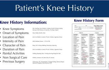
The Office Evaluation prior to surgery is used to review each patient’s symptoms, their knee history, their medical history, perform a physical exam, and to review imaging of the patient’s knee. This evaluation helps Dr. Buechel understand each patient’s situation, confirm that the physical findings align with the history and imaging, which allows Dr. Buechel to confirm the recommended procedure for each patient’s knee problem.
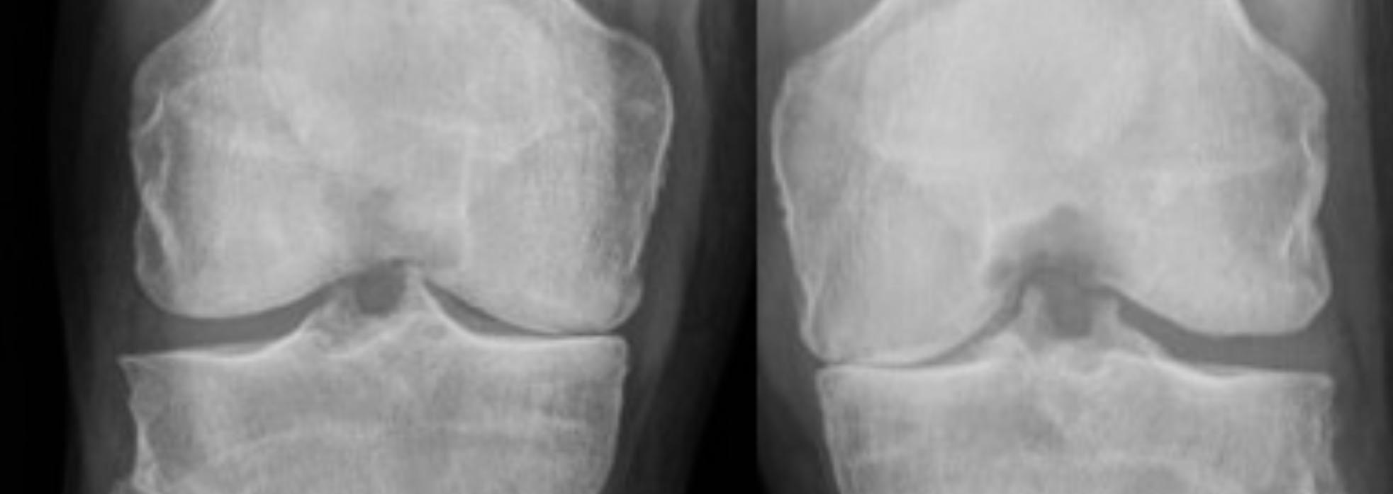
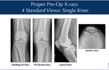
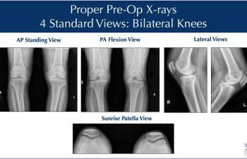
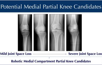
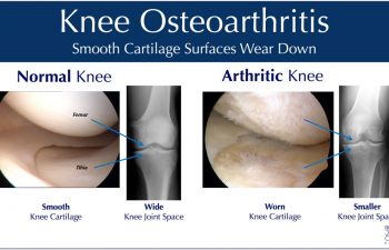
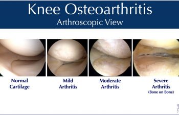
X-Rays for Evaluation of the Knee
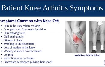
Patients obtain X-rays with 4 views of their knee to look for mild, moderate or severe joint space loss or destruction in one compartment from osteoarthritis, osteonecrosis or post-traumatic arthritis. If the x-rays and the exam are consistent with partial knee arthritis, and symptoms are present only in the affected side, then a patient may be a candidate for the Mako™ Robotic-Arm Assisted Partial Knee Resurfacing procedure. The range of medial compartment arthritis related joint space loss is shown, representing the potential candidates treatable with Mako™ Robotic PKA.
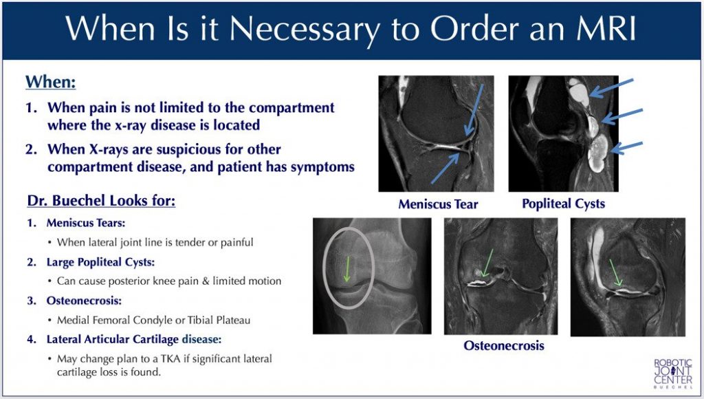
MRI Imaging of the Knee
An MRI is not needed on every patient. However, when the x-rays and the examination leave some doubt as to all the sources of pain, or whether another compartment involvement is too much for partial knee, an MRI is the best study to shed further light on the situation. The MRI provides more information on the cartilage condition, the ligament condition and the bone problems assisting in the decision-making process. This is generally the best overall second study, following the x-ray, if the decision is still questionable.
Clinical Indications & Contra-Indications for Mako™ Robotic Partial Knee Replacement
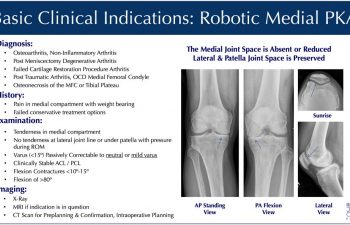
The indication to proceed with Robotic-Arm Assisted PKA is determined by the proper diagnosis, an appropriate history, Dr. Buechel’s examination that confirms the findings, and imaging studies that support these conclusions. The most common reason for knee replacement surgery is osteoarthritis. Other reasons include: arthritis after meniscectomy, post-traumatic arthritis, osteonecrosis, osteo-chondral defects, and failed cartilage restoration procedures.
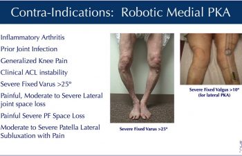
There are a few reasons that Dr. Buechel and other surgeons would recommend against a partial knee replacement which include generalized inflammatory arthritis conditions (i.e. rheumatoid arthritis), infection history in the knee, some severe deformities and ligament instabilities, and generalized pain throughout the knee not specific to one area.
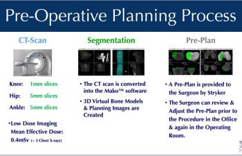
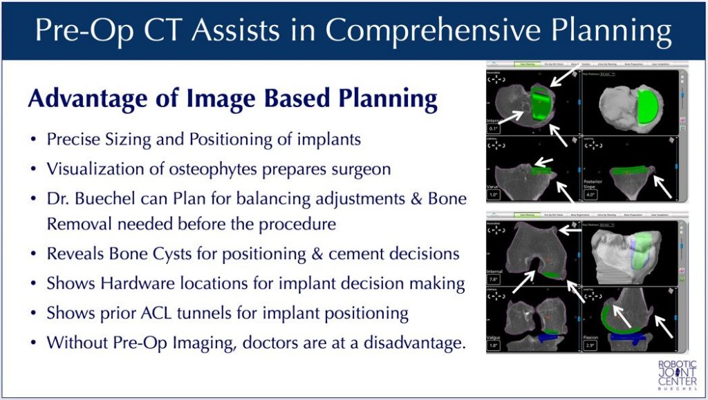
Pre-Operative Planning Process & Implant Planning
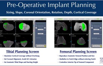
Pre-Planning is the process of preparing for the robotic knee procedure by obtaining precision 3D knee imaging data that is loaded into an advanced knee implant software platform. This allows the surgeon to position the virtual knee implants onto the patient specific knee bone models. Pre-planning allows the surgeon to plan the exact size, the implant depth, sagittal, coronal & transverse rotation, posterior slope, coronal plane alignment, and initial tracking position of the chosen implants, prior to the procedure.
As a critical part of the procedure, preoperative computer planning and evaluation makes a big difference in the efficiency and quality of the surgical procedure to follow, and the outcome. The preoperative CT scan provides enormous benefit to patients & Dr. Buechel. Once the CT data is loaded into the software, Dr. Buechel optimizes the patient’s implant positions based on each patient’s anatomy, with one millimeter and 1degree precision. The anatomic preoperative plan is optimized during surgery from motion data, tracking information, cartilage surface mapping, and gap analysis to optimize each patient’s individual proper tension and tracking pattern.
Value & Safety Provided by the Pre-Operative CT
The preoperative CT scans often reveal important information pre-operatively. Large bone spurs, often not seen with imageless systems, are important to know about preoperatively, as they influence implant positioning decisions during surgery, effecting proper joint tension. On the more advanced arthritis cases with lots of large bone spurs, the scans help tremendously in planning & ensuring their removal. The Pre-operative CT is also critical for implant planning in cases of osteonecrosis. In these cases, bone loss or bone death areas are properly assessed so Dr. Buechel can optimize the implant position to cover the bad areas and provide proper implant support. The CT scans sometimes reveal large bone cysts that may influence a surgeon’s decision to perform the procedure, or how to plan the procedure to ensure implant stability.
A concern patients have with CT scans is related to the radiation dosage and its potential detrimental effects. I have been concerned as well, so our radiologists have shared with us that the special protocol for knee scans delivers a mean effective dose of only ~0.4mSv, which is equivalent to 1.5 months of natural environmental radiation, or 3 x-rays. Therefore, I strongly feel the benefit outweighs the minimal risk.
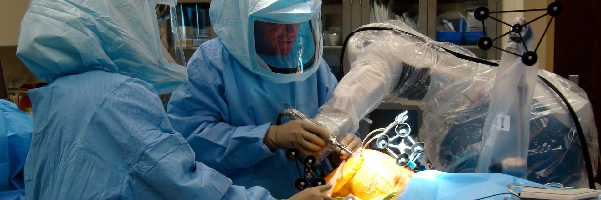
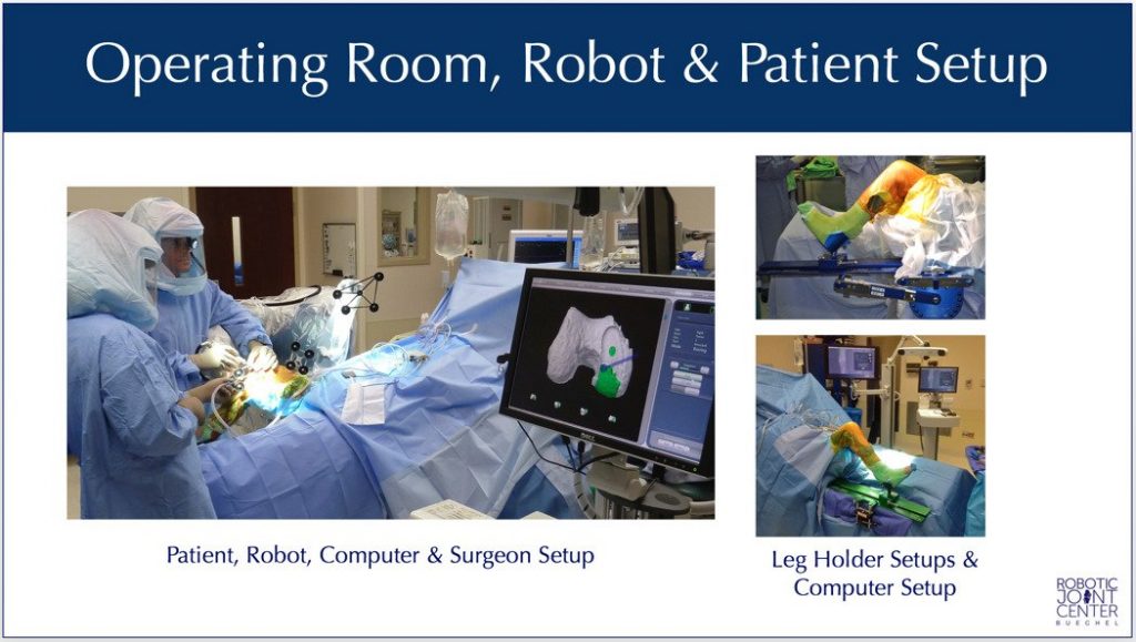
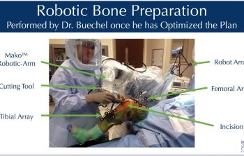
Patient & Robotic Operating Room Setup
Once the operating room is ready, the patient is brought into the operating room and properly positioned on a comfortable and well-padded operating table. Once the anesthesiologist has completed their preparation of the patient with either a regional or general anesthesia, the patient is sterilely prepared, padded and draped.
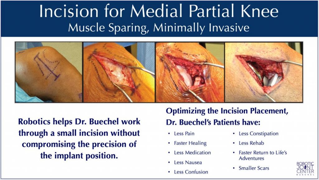
Incisions & Exposure
Dr. Buechel’s skin incisions are 2-3 inches for the medial partial knee procedures (3-4 inches for lateral side), no muscles are cut, no tourniquets are needed, and blood loss is minimal requiring no transfusions. Dr. Buechel has designed specialized tools to allow small incisions to be made while still allowing great access & visibility into the entire joint. These tools also aid in finely crafting the remaining bone edges and trimming tissues so they heal well, reducing the chance for impinging scar tissues that can complicate recovery. Advanced medicines are used to reduce blood loss during, and after surgery. Electro-cautery knives are used to minimize bleeding during the exposure. Skin adhesives and antibiotic coated barbed suture are used during closure on most patients to hold the tissue edges together better, reduce the risk of external contamination, while allowing patients to shower the next day.
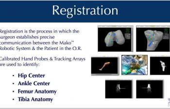
Tracking Arrays & Registration
During the procedure, Dr. Buechel places temporary infrared tracking devices called “Arrays”, on the femur and tibia. These allow the computer to “see” the bones moving in the operating room precisely, know the exact position of the robot with its cutting tool, and match the precise location of the bone of the patient to the 3D CT scan plan in the computer. This process is called “Registration”.
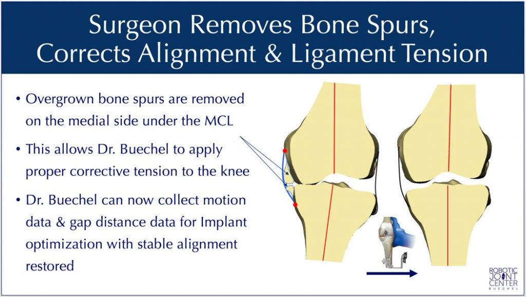
Bone Spur (Osteophyte) Removal in Preparation for Data Collection
Once the communication has been established by matching the patient to the CT data through the “Registration” process, any overgrown bone spurs can be removed. This is part of the preparation to properly re-tension the knee ligaments, individually re-align the knee, and then collect data to allow for optimal implant planning & positioning.
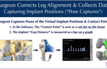
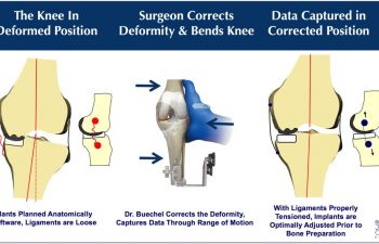
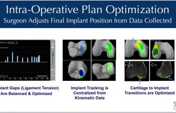
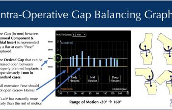
Data Collection (“Pose Captures”) & Adjustment of Implants (“Intra-Operative Planning”)
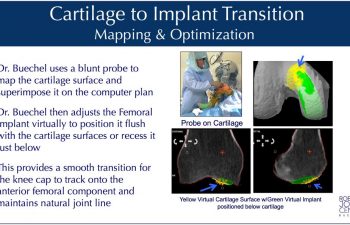
Once the software/patient/robot communication is setup in the operating room, Dr. Buechel bends the knee through a range of motion while he re-tensions the ligaments of the knee and optimizes the angular alignment for each patient. This allows the computer software to present the surgeon with tracking information, implant gap analysis, and cartilage surface mapping so the surgeon can make virtual adjustments of the implants, to optimize their position prior to robotic bone preparation.
Understanding how to optimize this data collection process and adjust for different deformities is where Dr. Buechel’s vast experience using the system makes a significant difference.
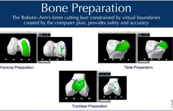
Robotic Bone Preparation
With the intraoperative plan optimized, Dr. Buechel guides a 6mm round cutting tool connected to the end of a robotic-arm across the bone surface, removing the planned volume of bone resection. The robotic system provides visual, audible and tactile feedback limiting the area of resection to only the planned surface, within a millimeter and one degree of the plan. The robotic system creates virtual boundaries like an “invisible wall”, safely and precisely limiting the movement of the cutting tool.
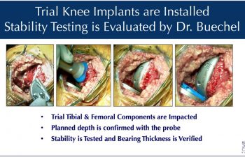
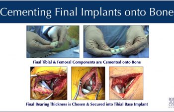
Implant Installation and Validation
Once the bone surface is prepared by the robotic cutting tool, Dr. Buechel then trims and prepares the remaining soft tissues in the joint space in preparation to install the trial implants, and then the final implants. Trial implants are installed without cement to test the tracking, tension and stability of the knee joint. The final implants are installed with the use of bone cement attaching them to the femoral and tibial bones. Probes are used to verify the precise seating of the implants to the desired location in the cement bed. A final check of stability is made with a trial bearing before the final bearing is chosen and locked into the tibial base implant.
Wound Closure & Healing Optimization
Dr. Buechel is focused on optimizing wound healing for all of his surgical patients. Therefore, he has recommendations on the entire process from before surgery, to after surgery to enhance wound healing and recovery.
Patients should optimize their preoperative nutrition and exercise, while eliminating or minimizing any risk factors for poor wound healing such as smoking, excessive alcohol intake, poor blood sugar control or excessive Body Mass Index (>40 BMI) which all can lead to greater complications after surgery. Immuno-nutrition products such as Ricochet drinks help optimize each patients’ nutritional status for elective surgery. Proper protein and nutritional intake before and after surgery has been shown to improve wound healing and reduce the risk of infection.
During the procedure, Dr. Buechel meticulously cares for the health of the skin and deep tissues throughout the procedure while exposing, retracting, and closing the knee joint, to optimize healing, minimize blood loss, and reduce the incidence of wound healing problems.
A cosmetically appealing scar is common after Dr. Buechel’s procedures because of the variety of techniques he employs during the surgery, and the carefully chosen products he uses for the closure and aftercare. The highest quality suture is used to hold the tissues longer and stronger, while providing anti-bacterial protection. Stratafix Plus barbed antimicrobial suture is our preferred suture.
The outer skin edges are sealed with skin adhesive that blocks out bacteria and provides a waterproof seal to allow showering after surgery. Dermabond skin adhesive is our preferred sealant.
High quality, hypoallergenic, waterproof dressings designed to be gentle on the skin are used to cover the surgical site, adding to the protection of each patients wound.
A Reparel Leg Sleeve is provided after surgery to help control tissue swelling and improve circulation during the early weeks of healing. The sleeves are comfortable, breathable, provide mild compression, are moisture wicking, and anti-microbial, and they use silicon grips at the top band to prevent slippage and folding. The material is composed of synthetic fibers that incorporate finely processed, non-metallic, elemental semi-conductor nanoparticles chosen specifically for their relative efficiency at transmitting energy when body heat contacts the material. The energy is then released as photons, “or particles of light” into the subcutaneous tissues and is thought to promote better cell metabolism /respiration and better circulation by its effects of accelerating the production of ATP in the cell mitochondria.
Cold Therapy wraps devices are critical in wound healing after surgery to reduce swelling and provide non-medication-based pain control. With the use of continuous flow cold therapy sleeves after surgery, patients can use cold as a mechanical way of reducing swelling and pain without the risks of ice packs. These devices allow for longer application without the risks of ice packs.
Dr. Buechel tries to optimize each individuals knee for healing by paying attention to the tissue handling during surgery, the closure technique and materials, and the post-operative wound environment. However, we all heal a little differently. Some people either genetically, due to their muscle tightness, or their adipose tissue thickness, form scar tissue thicker, faster, and tighter, and have wider scars. Some people form very little scar tissue, have more flexible tissue and have very fine line scars. Dr. Buechel uses a variety of techniques to optimize wound healing and appearance for each patient he operates on, hoping for an optimal final result.
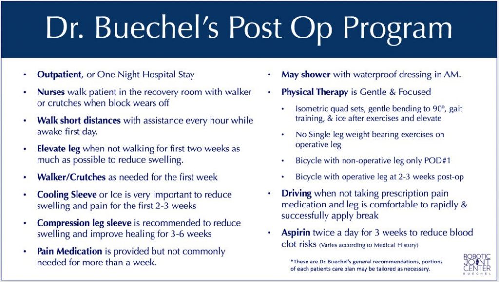
Immediate Post-Operative Recommendations
Patients will be discharged from the surgical facility once they are awake, alert, medically stable, and have had a chance to walk around the surgical facility with their assistive devices. Usually this is the same day. However, based on individual circumstances, if a patient needs more care or a longer stay, this can be arranged. Sterile dressings cover the knee with a mild compression wrap from mid-thigh to mid-tibia when patients leave the operating room.
A cooling knee sleeve device is recommended to be used when your arrival back at your home, or temporary housing location, for those traveling for their procedure. Patients are recommended to walk each hour for a short distance while awake with support at first, and then without support when they are feeling confident and stable. When resting and not walking during the first two weeks after surgery, the leg should be elevated to heart level or above for swelling reduction, and the cooling sleeve should be on the knee. Pain medications should be taken only as necessary, and is made less necessary by using the cooling device, and elevating the leg to reduce swelling.
Although blood clots are extremely rare with this procedure, the use of an anticoagulant medication is generally recommended for most patients for 3 weeks, with the specific choice determined for each patient based on their risk factors. Ankle pumping exercises and frequent short walks daily beginning the day of surgery significantly reduces the risk of blood clots (DVT). A compression stocking is recommended to help control swelling and a specific leg healing sleeve that Dr. Buechel has found to be comfortable and promote healthy healing will be provided in the office, after the procedure.
Patients return to the office the day after their procedure for a checkup at which time their knee sleeve is provided, the therapy is instituted, x-rays are reviewed and all concerns are addressed.
Rehabilitation
A post procedure exercise program is recommended to optimize your outcomes and optimize your quality of life moving into the future. Patients can begin a progressive exercise program the day of surgery. Dr. Buechel’s comprehensive Mako™ Robotic-Arm Assisted Partial Knee Replacement Program is designed to enhance patient outcomes, allowing patients the greatest chance of success with the least risk of complications. Local therapists who work with Dr. Buechel are recommended for patients so they can begin their exercise programs immediately. Those that will be returning to their home towns can take their initial training program plan with them, and continue back at home when they arrive.
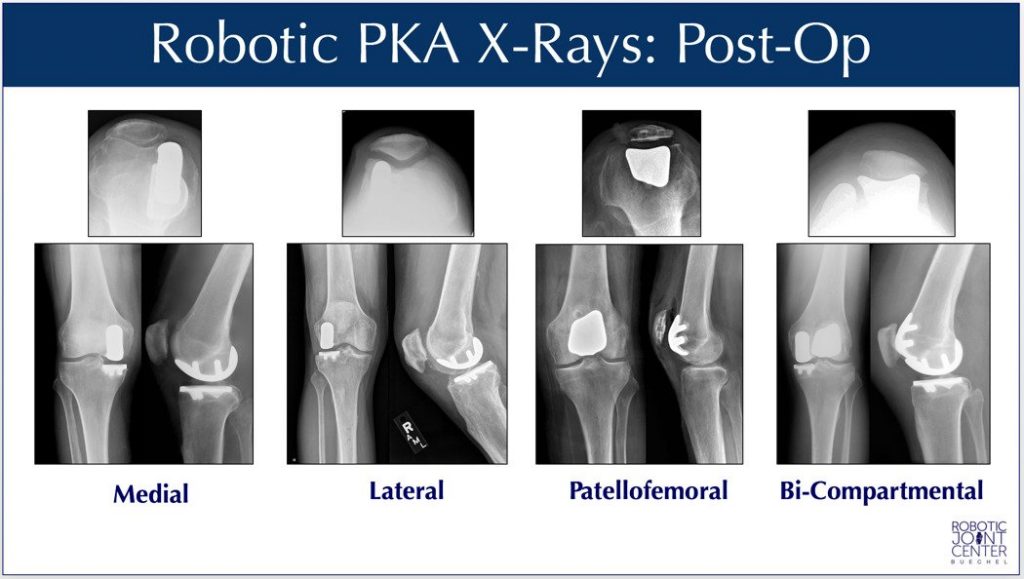
X-Rays of Dr. Buechel’s Mako™ Robotic-Arm Assisted Partial Knee Replacements
Patients have options to have the individual compartments of their knee replaced (or resurfaced) using the precision Mako™ Robotic System. The compartments that can be treated are called the Medial, Lateral, and Patellofemoral. The Medial and Patellofemoral can be performed together at the same time for those that have symptoms in both compartments but wish to retain their natural cruciate ligaments and healthy lateral compartment.
X-Rays of Dr. Frederick Buechel, Jr. MD’s Robotic-Arm Assisted Partial Knee Replacements showing all “FDA Approved” options.
Written by: Dr. Frederick Buechel, Jr.
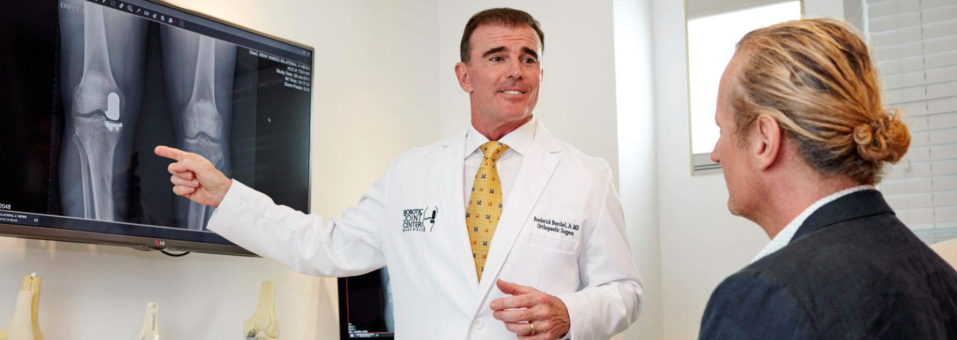
Dr. Buechel is a Stryker certified international instructor and an expert in the field of Robotic-Arm Assisted Knee & Hip Replacement.
Dr. Buechel performs Outpatient Robotic Partial Knee replacement surgery, and Outpatient or short stay Robotic Total Knee replacement in Naples, Florida. Internationally, Dr. Buechel performs Robotic Knee and Hip replacement in Taipei, Taiwan, at Taipei Postal Hospital.